
Table of contents
Cranium - Morphological regions (Clypeus - Face - Frons - Vertex - Occiput - Compound eye - Gena - Postgena) - Ocelli - Chaetotaxy - Bibliography - Web resources)The head of Diptera is well differentiated and evident, separated from the thorax by a short membranous region named neck. The shape is subglobose, with a possible further development in width or length, sightly accentuated. Only few groups have a very long head, such some Tipulomorpha. Regarding the relative position, the condition most frequent is the axis more or less perpendicular to the body (hypognathous head), so the mouthparts are directed downwards, compared to the horizontal posture assumed by the insect.
Such as all Insects, the head was derived from the deeply modification and fusion of first six segments, named cephalic somites and distinguished into preoral and postoral or gnathal. It is morphologically divided into several regions and is provided of two types of appendages, the antennae, paired and symmetrical, and the gnathites, these last components of the ensemble of mouthparts.
Inside the head there are the cerebrum, the subesophageal ganglion, most of the dorsal sympathetic nervous system, the end of aorta and the cephalic portions of tracheal and muscular systems.
Cranium
As in most Insects, the cranium appears like a capsule more or less rigid, formed by the exoskeleton. It is open in the lower and posterior sides (oral and occipital foramens).
Inside the cephalic cavity, the exoskeleton is invaginated and forms the part more complex of endoskeleton, named tentorium. These invaginations may be correspond to small symmetrical pits in the outer surface (tentorial pits), on the facial and occipital regions. In the primitive structure of tentorium, there are two anterior arms (pretentoria), that arise from the facial region, and two posterior arms (metatentoria), which arise from the postgenae; these arms are joined in the tentorial bridge and give to the structure a X shape. There are also two dorsal arms (supertentoria) which arise from the anterior arms and are directed upward, toward the front, without reaching the inner surface of exoskeleton. The tentorium of Dipters has a marked reduction, compared with the primitive structure; this reduction concerns in particular the development of the tentorial bridge, which is usually incomplete or absent in most of the order.
The posterior tentorial pits are generally barely visible, under and lateral to occipital foramen, in the posterior side of head. The anterior tentorial pits are barely visible or absent in most of Schizophora, while they are well developed in certain families of Nematocera and in Tabanomorpha.
Morphological regions
In the head of most Dipters we distinguish, with various extensions and forms, the follow regions:
- unpaired and median regions: from anterior side to posterior, there are the clypeus, the face, the frons, the vertex and the occiput;
- lateral regions, paired and symmetrical: from top to bottom, on each side there are the compound eye, the gena, and posteriorly the postgena.


1: ocellar triangle; 2: vertex; 3: frontal vitta; 4: frontorbital plate; 5: compound eye; 6: lunule; 7: frontal suture; 8: parafacialia; 9: facial ridge; 10: vibrissal angle; 11: face; 12: clypeus; 13: labium; 14: labellum; 15: clypeal membrane; 16: maxillary palp; 17: frontoclypeal membrane; 18: frontogenal suture; 19: antenna; 20: carina; 21: ocellus; 22: prementum; 23: proboscis; 24: peristoma; 25: genal dilation; 26: postgena; 27: gena; 28: genal groove; 29: occiput.
Author: Giancarlo Dessì
(License: Creative Commons BY-NC-SA)
Clypeus
The clypeus is a sclerite placed in the lower part of head, under the face and upper the labrum; extensions and forms are diversified among the order. The inside gives connection to cibarial dilator muscles and the region has a particular develop in ematophagous groups.
In most lower Diptera (Nematocera e Orthorrhapha), the clypeus appears as a shield more or less wide, with various shapes, connected to the face by the frontoclypeal suture. In the Tipulidae and other genera of Tipuloidea, the clypeus is particulary elongate and it forms, with the genae, a prolongation of head ("nasus") bearing the mouthparts at the tip. The distinction between the clypeus and the adjacent regions, particularly the genae and the face, is not clearly defined. In the Cyclorrhapha, the clypeus appear as a small sclerite U-shaped, visible at the base of the mouthparts, and it connects to the face by a frontoclypeal membrana, wide and flexible. These marked morphostructural differences reflect in the appearance of head in the frontal view:
- in the lower Diptera, the clypeal region appears as an area more or less bounded, placed in lower part of anterior side of head;
- in the Tipulidae and part of Tipuloidea it appears as an area not bounded, placed on the anterior prolongation of head;
- in the Cyclorrhapha it appears as part of "proboscis" and is reduced to a small area at the base of anterior side.
The distal edge of clypeus is connected to the labrum by a true articulation or a membrana (clypeal membrana). In some groups (e.g. Blephariceridae), the clypeus is divided into a proximal (postclypeus) and a distal (anteclypeus) regions.
Face
The face, or prefrons, is a median region which extend, roughly, between the anterior edges of compound eyes, the insertion of antennae and the upper edge of the clypeus or the frontoclypeal membrana. Shape and width of this area vary depending of systematic groups, but, in general there are marked differences between the lower Diptera and the Cyclorrhapha.
Morphology of the face in lower Diptera
In most lower Dipters (Nematocera, Orthorrhapha, and Aschiza), the face is not well developed and the limits are undefined. This condition occurs particularly in ematophagous forms, where the wide development of clypeus is achieved at the expense of face. In some Orthorrhapha (e.g. Asilidae) and in few of Nematocera families, the face is wider. In the Asilids, the face is strongly convex and is covered by a thick tuft of bristles (mystax).
Morphology of the face in higher Diptera
In the Schizophora, the face is well developed and has a complex structure related to the presence of the ptilinal suture and the frontogenal sutures. In these dipters, the area between the anterior edges of eyes is composed of three sclerites: the median, called facial, and two lateral, paired and symmetrical, named parafacialia. The face appears as a subtriangular area, wide in the lower, more or less narrow in the upper part. It is crossed by the frontal suture or ptilinal suture, derived from the invagination of the ptilinum. The ptilinal suture runs dorsally from the sides of the lower part of the face, drawing an inverted U-like or V-like line. The suture separates the facial plate laterally from the parafacialia and dorsally from the frons. At the sides of the facial plate there are other two symmetrical sutures (frontogenal sutures), placed medial to ptilinal suture, which run vertically from the insertion of vibrissae to the anterior tentorial pits, near the insertion of antennae.
The parafacialia, paired and symmetrical, are between the anterior edge of the eye and the ptilinal suture and appear more or less narrow and elongated. These sclerites are connected with the frontorbital plates, dorsally, and the genae, ventrally. Their surface may be bare or setose.
The face plate shows some morphological characters useful for taxonomic determination. The two narrow strips between the ptilinal and frontogenal sutures are named facial ridges or vibrissal ridges. The ventral end of the faccial ridge, named vibrissal angle, appear as a relief more or less prominent that often bears a thick and stout bristle, named vibrissa. Except to vibrissae, present in several families of Cyclorrhaphous dipters, the facial ridges are usually bare, but sometimes there are other setae, named supravibrissal bristles (e.g. Tachinidae).
The upper side of facial plate bears the insertion of antennae, which are more or less close in the Cyclorrhaphous. The small area strongly sclerotized present above the insertion of antennae and bounded from the dorsal part of the ptilinal suture is called lunule. The lunule is typical of Schizophora, however a similar structure is present also in some Aschiza. In several Schizophora, a median reliev called carina may arise from the lunule and it runs vertically to base of the facial plate. The carina, when present, separates two grooves, called antennal grooves, because they receive the antennae.
Frons
The frons or postfrons is the median region between the dorsal edges of eyes and the vertex; the lower limit is represented by the insertion of antennae in the lower Dipters and by the ptilinal suture in the Cyclorrhapha. Therefore, the name "frons" is used, about the Diptera, with a sense closer than the one recurrent for other orders, where usually the frons is the entire area between the eyes, the genae, the vertex, and the frontoclypeal suture.
The width of frons depends on the shape of the head and, especially, on the development of eyes. In particular, the frons is virtually abstent or very reduced in the males with holoptic head, because the conjunction of eyes occurs in the frontal region.
Also for the frons we should distinguish between lower and higher Dipters cause of strong morphological differences. In the first group includes Nematocerous and Orthorrhaphous Dipters and the Aschiza, the second one includes the Schizophora.
Morphology of the frons in lower Diptera
In lower Diptera (Nematocerous, lower Brachycerous and lower Cyclorrhaphous), the frons appears as a region with uniform look, except for a few groups that may show some morphological details (e.g. the calli of Tabanidae).
Morphology of the frons in higher Diptera
In Schizophora, the morphology of the frons, especially the chaetotaxy, is a character of great taxonomic importance. Except for species with holoptic head, the frons is usually wide and divided into three areas, one unpaired and median and two paired and symmetrical.
The median area is called frontal vitta, interfrons, mesofrons. It is a weakly sclerotize region, subrectangular or trapezoidal, which extend from the ocellar triangle up the lunule, sometimes spreading up the entire width of the frons in the ventral side.
The lateral areas, paired and symmetrical, are called frontorbital plates or, rarely, orbits or parafrontal plates. In general, as a primitive condition, each frontorbital plate is composed of a single sclerite, that extends between the dorsal edge of eye and the frontal vitta, from the vertex to the upper limit of parafacialia. Apomorphic conditions may give different development of frontorbital plates. The most important are the following:
- reduction to the obliteration in the ventral side (e.g. Heleomyzidae): the frontorbital plate is ventrally broken by the enlargin of frontal vitta and it is reduced to a narrow area parallel to the dorsal edge of eye, in the upper portion of the frons;
- division into two sclerites, more or less separated (e.g. Tephritidae): the frontorbital area is composed of two distinct sclerites, named frontal plate and orbital plate. The first one is medial and is placed on the dorsal part of the frons, arising from the vertex; the second one is lateral, is parallel to the margin of eye and reaches the upper limit of parafacialia;
- division into two indistinguishable sclerites: this condition occurs in most Calyptratae. As before, the area is composed of two sclerites, but their fusion does not allow the morphological distinction.
Vertex
The vertex is not a true region, cause of the absence of clear limits: it corresponds to the top of head, between the eyes, the frons, and the occiput and has a natural continuity with these last two regions. In Nematocera, the vertex is integral part of the frons and forms the dorsal portion (McAlpine, 1981), while in Brachycera, especially in the Cyclorrhapha, the vertex is more easily identifiable for the position of some morphological elements.
In the vertex there is the ocellar triangle, although this structure extends also in the frons. The ocellar triangle is a plate more or less differentiated, often most raised, including the ocelli in the edges or slightly inside these. The ocelli are placed on a triangular profile, with the median ocellus more anterior than the lateral. At the sides of the ocellar triangle, in the space between the lateral ocelli and the top of eyes, there is the dorsal part of the orbital or frontorbital plates.
The broadness of vertex depends on the development of eyes and the shape of the head. In flies with dichoptic and broad head, the vertex is wide, while in the eyes with holoptic and globose head, the vertex is virtually absent and is represented only by the ocellar triangle.
The region has a great importance in the description of cyclorrhaphous dipters, because their taxonomic determination is often referred to chaetotaxy of the vertex and the frons.
Occiput
The occiput is the region between the posterior edges of eyes, the vertex, and the postgenae in the ventral side. It occupies most of the back of head and the limits between the occiput and the postgenae is marked by the posterior tentorial pits and the occipital foramen.
The conformation of this region depends essentially on the shape the head. In most Nematocera, characterized by the head subglobose, the occiput has a convex profile, while in most Brachycera is flat or concave.
The morphological structure and the consequent appearance of occiput vary considerably between the different systematics group, achieving complexity in most Brachycerous. Except for some Orthorrhapha, in these flies, the occiput is composed of several sclerites. Above the occipital foramen there is the median occipital sclerite, that occupies the entire central band from the occipital foramen to the ocellar triangle. At the sides of median there are two symmetrical sclerites, than have not a specific name. These sclerites are surrounded by the median sclerite, the posterior edge of eyes, and the posterior tentorial pits. Around the occipital foramen there is a narrow sclerotized rim, called postoccipitum. The lower limit of occiput is not well defined and is marked only by posterior tentorial pits, that separate the occiput from the postgenae.
As other regions described above, also the occiput is related to a specific chaetotaxy, useful sometimes for taxonomic purposes.
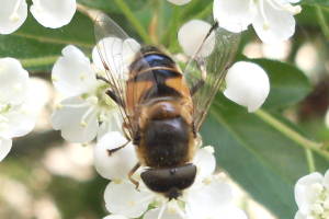
Compound eye

Author: Louisa Howard (Dartmouth College)
Resized from the original picture
(License: Public Domain)
In most of Dipters, the compound eyes are the morphological region most evident, because they may occupy most of head surface from the dorsal view and, above all, from the lateral view. In some groups, the eyes may also be reduced until the total absence (e.g. Braulidae), condition that occurs sometimes in parasitoid, commensal, or hypogeous forms.
From anatomical and physiological point of view, the compound eye is the organ that receives the light stimuli and converts them into neurochemical signals. This function, in most flies plays an important role. The eye is structurally composed of a geometric association of a large number of primatic and pyramidal elementary units, called ommatidia; their hexagonal section narrows descending from top to bottom. Each ommatidia is composed of a lens (corneal lens), a reflective body (crystalline cone), and a system of neurosensorial cells (retinula cells). Outside, the eye's surface appears subspheroidal and multifaceted; this is han effect due to the regular geometrical arrangement of ommatidia, wich are visible only corneas, being tightly packed to each other. Among the ommatidia a regular short hairs or setulae may emerge among the ommatidia. The presence and number of these macrotrichia is an useful in taxonomic descriptions. The number of ommatidia can be very large: the eye of the housefly (Musca domestica Linnaeus), for example, has over than 4,000 ommatidia (Tremblay, 1991).
From morphological point of view, the most important character is the the relative position of the eyes. Two basic conditions occur among the Diptera:
- holoptic head: the eyes extend dorsally to join in the frontal region, keeping space only to ocellar triangle;
- dichoptic head: the eyes are well separated by large vertex and frons.
The holoptic head occur over all in the males of many families, both in Nematocera and Brachycera. However there are families where the holoptic head is a character present in both sexes in all systematic groups, both Nematocerous (e.g. Thaumaleidae) and Brachycerous (e.g. Acroceridae). Many Authors agree that the condition of holoptic head is related to ethological contexts: in fact this character occurs in males of taxa which form dancing swarms or have behaviors based on the courtship in flight or the territorial control (McAlpine & Munroe, 1968; McAlpine, 1981; Merz & Haenni, 2000). However, the holoptic head also appears in many groups that do not show thes behaviors. Another character that occurs in some nematocerous families is the ocular bridge (e.g. Sciaridae, Scatopsidae): the eyed are fused in the vertex by a bridge of ommatidia. In contrast, other groups show the suddivisione of each eye in two separate and morphologically different areas (e.g. Bibionidae, Perissommatidae). Finally, other groups may have the ommatidia differentiated into two types, the one normal and the other large (e.g. Bibionidae, Blephariceridae, Simuliidae, etc.). An important fact is that the pigmentation is generally lost after the insect death.
Gena
The gena is a paired and simmetrical sclerite that occupies the side of head below the eye. Extension, appearance, and connection with other regions change in different systematic groups. Generally is included between the lower edge of the eye, the postgena, the peristomal margin, and the face. According to the Manual of Nearctic Diptera, the genal region is divided into two sclerites by the subgenal suture. This suture arises from the anterior tentorial pit and runs below the eye. The dorsal sclerite, that occupies the most of genal region, is called "gena" sensu stricto, while the ventral one, very narrow, is called subgena (McAlpine, 1981). This distinction does not appear in other works; moreover, the subareas treated by McAlpine are strictly related and not easily discernible.
In lower Diptera, that have a simple morphology of frontal and facial regions, the gena connect dorsally and anteriorly to the frontorbital plate. In tipuloid Nematocera, the genae are porrect and fused with the clypeus, forming a characteristic prolongation of the head called nasus. In Cyclorrhapha the connection with anterior regions is more complicated: the anterior margins of genae are connected to the facial ridges at the lower side and to parafacialia at the dorsal side. The posterior margins are connected to the postgenae, the sclerites which correspond to the gula of other insects. The limit between the gena and the postgena is non well defined, due to the merger of these sclerites.
Other morphological characters of this region are typical of several Cyclorrhapha and are called genal groove and genal dilation. The genal groove is a depression weakly sclerotized, placed near the limit between the gena and the parafacialia and is usually bare; it is large in the Calyptratae. Below the genal groove and behind the vibrissal angle there is the genal dilation, which unlike the groove is strongly sclerotized and usually pilose.
Postgena
The postgena is a small region, paired and simmetrical, placed in the lower part of the posterior side of head. It is laterally bounded by the gena, dorsally by the occipitum, ventrally by the hypostoma. The limits of postenae are not well defined, because of the fusione of postgenae with the genae and the occiput. As marked limits there are only the posterior tentorial pits that separate the occiput from the house.
In the middle, ventrally to occipital foramen and the tentorial pits, the limit between the two regions is not defined cause the fusion of this sclerites. The transitional area is called hypostomal bridge or pseudogula and is present in most Diptera except for some Nematocerous families.
Ocelli
Most flies have three ocelli arranged at the vertices of a triangle, with a median ocellus and two posterior and lateral. Some groups have less than three ocelli, due to lacking of the median or the lateral or the total absence. These conditions occur over all in Nematocera, but the absence of ocelli may be occur in Brachycera also (e.g. Braulidae).
In Cyclorrhapha, the ocelli are arranged on a plate, called ocellar triangle or ocellar plate, in the vertex. The ocellar triangle is generally well distinct from surrounding areas, sometimes prominent. In some groups, it is very larger (e.g. Chloropidae).
In lower Diptera the ocelli are generally arranged on a triangular perimeter on the vertex. This perimeter may be undifferentiated or it is borne by a protuberance called ocellar tubercle. In some families may be occur other unusual positions, such as Nymphomyiidae, which have only two ocelli behind the eyes.
Chaetotaxy
The chaetotaxy of head is one of the main characters treated in the systematic determination for the Cyclorrhapha. In the literature, the terminology is not uniform: Steyskal (1976) revised the nomenclature concerning the chaetotaxy of occiput and vertex and introduced some inconsistencies with the terminology of previous works. The terminology sensu Steyskal was adopted also by McAlpine et al. in the three volumes of the Manual of Nearctic Diptera (1981, 1987, 1989) and, partially, in next works. However, we must add, to incongruences caused by different criteria, also different point of view concernig the homology. Among these there is, for example, the dispute between the Canadian James F. McAlpine (1989) and the Australian David K. McAlpine (2007), concerning the origin of some bristles in the upper occipital region in the Canacidae. So, the terminology may be confused cause different terms used in different works. This website refers to terminology proposed in the Manual of Nearctic Diptera (McAlpine, 1981), but will cite, if necessary, the main discrepancies found in the literature.


gn: genal; f orb: frontorbital; fr: interfrontal; oc: ocellar; pgn: postgenal; po: postocellar; poc: postocular; pvt: paravertical; sp v: supravibrissal; st b: subvibrissal; vb: vibrissae; vt e: outer vertical; vt i: inner vertical.
Author: Giancarlo Dessì
(License: Creative Commons BY-NC-SA)
The frons, like the vertex, is one of regions that are mainly treated to characterize the chaetotaxy of head. Sometimes the bristles (or other macrotrichia) of the frons are called frontorbital as generic name, but the strict meaning of this name refers only to bristles aligned on the frontorbital plates. According to the Manual of Nearctic Diptera (McAlpine, 1981) and the Manual of Palaearctic Diptera (Merz & Haenni, 2000), bristles and other macrotrichia of the frontal region have this names:
- interfrontal: borne by the frontal vitta, they differ in number across the various families and sometimes are absent. Interfrontal bristles are usually weak;
- frontal: they are present in a few families only and are borne by the frontal plates when these sclerites are distinct from the orbital plates;
- orbital: like as the previous type, they are present in a few families and are borne by the orbital plates when distinct from the frontal plates;
- frontorbital: this type represents the most frequent condition. The bristles are aligned on the frontorbital plates between the frontal vitta and the eyes.
The orbital bristles are often identified by the position along the longitudinal axis. So they are distinct into lower orbital (onward) and upper orbital (behind). An important character of these bristles is their inclination, that may be lateroclinate (outward), medioclinate or inclinate (inwards), reclinate (backwards), and proclinate (forward). Other characters are the number and the size.
On the vertex there are usually two pairs of bristles and often are strong and elongate. Their position is lateral to the ocellar triangle and they are transversely aligned. This bristles are called vertical and are distinct into outer and inner vertical (McAlpine, 1981) or into lateral and medial vertical (Merz & Haenni, 2000). The size and the inclination may be considered in descriptions.
From the ocellar triangle two types of bristle are usually treated. The first pair, called ocellar, is composed by two symmetrical bristles usually inserted on lateral margins of the triangle, behind the median ocellus. The second pair is composed by two symmetrical bristles inserted on posterior margin of the triangle, between the lateral ocelli. These last bristles was called postvertical by Osten-Sacken (1884) and after by Hendel (1916). This name was widely used in the literature until '70 years, but Steyskal (1976) proposed postocellar as new name. The term of Steyskal is now used in most the recent literature, because it was adopted by the Manual of Nearctic Diptera (McAlpine, 1981; McAlpine et al., 1987; McAlpine, 1989) and the Manual of Palaearctic Diptera (Papp et al., 1998; Merz & Haenni, 2000), but some Authors use still the old name postvertical (McAlpine, 2007). Rarely, an additional pair, named medial postocellar may occur between the postocellar bristles (Merz & Haenni, 2000). Postocellar bristles have a great importance in taxonomy, because thei presence, size, and inclination may be determinant in the classification of Acalyptratae. The adjectives divergent, parallel, convergent and cruciate are used to describe the inclination of postocellar bristles.
In the vertical region may be also present other 1-2 pairs behind the vertical bristles or sometimes behind the ocellar triangle (e.g. Canacidae). The terminology of this macrotrichia is also confused and controversial. Hendel (1928) called them as inner occipital and Steyskal (1976) paravertical. The terminology of Steyskal was adopted by McAlpine (1981) in the Manual of Nearctic Diptera, but, in second volume, Vockeroth (1998) described the Tethinidae (=Canacidae) referring to terminology sensu Hendel. In the Manual of Palaearctic Diptera, Merz & Haenni (2000) refer this macrotrichia as paravertical and the same terminology is applied in the description of all Cyclorrhaphous families that have these bristles (Chandler & Shatalkin, 1998; McAlpine & Shatalkin, 1998; Munari, 1998; Rognes, 1998).
The occipital region have macrotrichia in most families, but this character has not a great importance and in general Authors merely report the presence. Macrotrichia present in this region are distinct into postocular, occipital and supracervical. The postoculars are bristles more or less developed and aligned in one or more rows parallel to posterior margins of eyes; the number of setae on each row is various, but is generally conspicuous. The occipitals, not to be confused with the inner occipitals sensu Hendel, are thick and scattered bristles medial to postoculars. Their distribution is usually irregular, but in some families may be regulary aligned. The supracervicals are short hairs of the median occipital sclerite, above the occipital foramen. Usually they have not importance.
The face is a region generally bare, but often in this region there are the vibrissae, two strong bristles inserted on the vibrissal angles, near the ventral extreme of frontogenal suture. On the facial ridges may be present other bristles similar to vibrissae and called supravibrissal bristles.
The genal region has usually macrotrichia of minor importance for taxonomic purposes and the descriptions refer only about their presence. The bristles of this region are called genal bristles and may vary in number and distribution. Among the genal bristles a few anterior, inserted near the vibrissal angle, may be longer and stronger than the posterior, similar to vibrissae. So, they are called subvibrissal bristles.
The postgenae have usually many hairs or setulae, called postgenal. They vary in number but in the taxonomic description is reported only the presence.
The chaetotaxy sensu stricto refers mainly to macrotrichia listed above, however there are hair or bristles also in other regions of the head. In particular may be reported:
- the pubescence of antennae and possibly the presence, number, and position of macrotrichia in proxymal articles;
- the presence of hairs in the compound eyes with regular distribution;
- the presence of hairs on the mouthparts, especially on the maxillary palps.
Bibliography
- Cerretti, P. (2010) Terminologia morfologica: 8-11. In I tachinidi della fauna italiana (Diptera Tachinidae), con chiave interattiva dei generi ovest-paleartici. Vol. II. Atlante iconografico, Work cited.
- Chandler, P.J. & Shatalkin, A.I. (1998) 3.3. Family Platypezidae: 27-50. In Papp, L. & Darvas, B. (eds.). Contributions to a Manual of Palaearctic Diptera. Volume 3. Higher Brachycera, Work cited.
- Hendel, F.G. (1928) Zweiflügler oder Diptera. II. Allgemeiner Teil, Work cited.
- Matile, L. (1993) Morphologie des Diptères: 54-96. In Diptères d'Europe Occidentale. Tome I, Work cited.
- McAlpine, D.K. & Shatalkin, A.I. (1998) 3.8. Family Pseudopomyzidae: 155-164. In Papp, L. & Darvas, B. (eds.) Contributions to a Manual of Palaearctic Diptera. Volume 3. Higher Brachycera, Work cited.
- McAlpine, D.K. (2007) The Surge Flies (Diptera: Canacidae: Zaleinae) of Australasia and notes on Tethinid-Canacid morphology and relationships, Work cited.
- McAlpine, J.F. & Munroe, D.D. (1968) Swarming of Lonchaeid flies and other insects, with descriptions of four new species of Lonchaeidae (Diptera), Work cited.
- McAlpine, J.F. (1981) Morphology and terminology - Adults: 9-63. In McAlpine, J.F.; Peterson, B.V.; Shewell, G.E.; Teskey, H.J.; Vockeroth, J.R. & Wood, D.M. (eds.) Manual of Nearctic Diptera. Volume 1, Work cited.
- McAlpine, J.F.; Peterson, B.V.; Shewell, G.E.; Teskey, H.J.; Vockeroth, J.R. & Wood, D.M. (1987) Manual of Nearctic Diptera. Volume 2, Work cited.
- McAlpine, J.F. (1989) Phylogeny and classification of the Muscomorpha: 1397-1518. In Borkent, A.; McAlpine, J.F.; Wood, D.M. & Woodley, N.E. (eds.) Manual of Nearctic Diptera. Volume 3, Work cited.
- Merz, B. & Haenni, J.P. (2000) 1.1. Morphology and terminology of adult Diptera (other than terminalia): 21-51. In Papp, L. & Darvas, B. (eds.) Contributions to a Manual of Palaearctic Diptera. Volume 1. General and Applied Dipterology, Work cited.
- Munari, L. (1998) 3.19. Family Tethinidae: 243-250. In Papp, L. & Darvas, B. (eds.) Contributions to a Manual of Palaearctic Diptera. Volume 3. Higher Brachycera, Work cited.
- Osten-Sacken, C.R. (1884) An essay on comparative chaetotaxy, or the arrangement of characteristic bristles of Diptera, Work cited.
- Papp, L. & Darvas, B. (1998) Contributions to a Manual of Palaearctic Diptera. Volume 3. Higher Brachycera, Work cited.
- Rognes, K. (1998) 3.51. Family Calliphoridae: 617-648. In Papp, L. & Darvas, B. (eds.) Contributions to a Manual of Palaearctic Diptera. Volume 3. Higher Brachycera, Work cited.
- Steyskal, G.C. (1976) The terminology of bristles on the upper back of the head in the higher Diptera, Work cited.
- Tremblay, E. (1991) Ordine Diptera (Ditteri): 11-20. In Entomologia applicata. Volume III Parte I, Work cited.
- Vockeroth, J.R. (1987) Tethinidae: 1073-1078. In McAlpine, J.F.; Peterson, B.V.; Shewell, G.E; Teskey, H.J.; Vockeroth, J.R. & Wood, D.M. (eds.) Manual of Nearctic Diptera. Volume 2, Work cited.
Web resources
- Yeates, D.K.; Hastings, A.; Hamilton, J.; Colless, D.H.; Lambkin, C.L.; Bickel, D.J.; McAlpine, D.K.; Schneider, M.A.; Daniels, G. & Cranston, P.S. Anatomical Atlas of Flies. In CSIRO Entomology. CSIRO, Commonwealth Scientific and Industrial Research Organisation. Last access: 28 May 2019.

Creative Commons BY-NC-SA 3.0 Unported License
(BY: Attribution - NC: Noncommercial - SA: Share Alike).